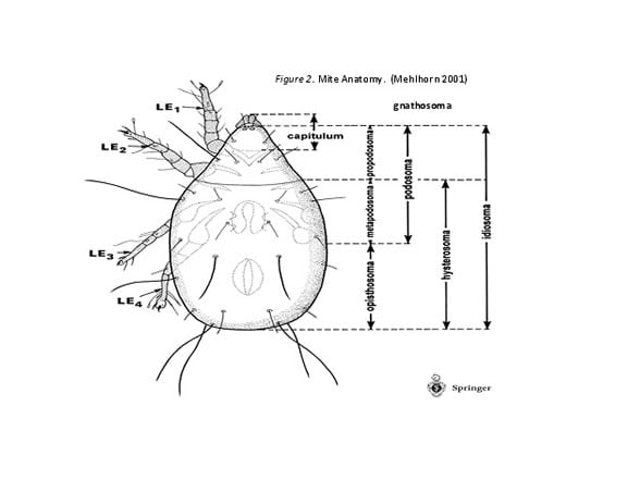
The anatomies of acarines, as shown in Figure 2, are fairly similar. The capitulum is the head and the idiosoma is the body. The idiosoma is composed of the propodosoma, metapodosoma, and opisthosoma. The segmentation that divides the propodosoma from the hysterosoma is the hallmark of Arachnida (Savory 1964). It is important to note that in Acari there is no division between the metapodosoma (thorax) and the opisthosoma (abdomen). This makes the body rounded (Mehlhorn 2001). Each leg (LE) is numbered starting from the head.
Adult mites have eight (four pairs of) legs versus their 6-legged insect cousins from the subphylum Hexapoda (six legs in Greek). The top portion of the capitulum, the gnathosoma, has two additional pairs of short appendages (one of which are the chelicerae mentioned earlier) that have evolved for feeding, sensing, and reproducing. Also, they don’t have antennae or wings like insects, but they do have hair-like bristles called setae.
Figure 2. Mite Anatomy. (Mehlhorn 2001)

The Northern Fowl Mite or O. sylviarum
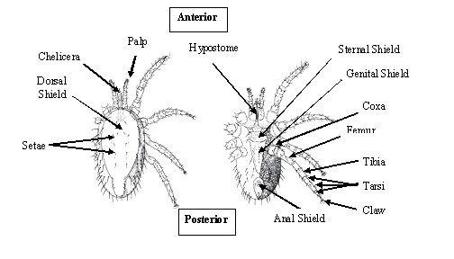
Figure 3 below shows a female of the species in which I have labeled the major parts. The relatively short pincer-like chelicerae (pair of chelicera) are the biting jaws that pierce the skin and suck the blood. These have chelae or pincers that are toothless (Lucky et al. 2001). On either side are palps (“a touching” in Latin) or elongated, segmented appendages near the mouth, functions of which include sensation and feeding. The hypostome is the lower lip.
Figure 3. Parts of a female O. sylviarum. Dorsal view on the left and Ventral on the Right (Drawings by E. W. Baker National Pest Control Association Inc.)

There are four shields or plates: the dorsal, sternal, genital, and anal. Proctor and Knee (2006) note a one-piece dorsal shield and point out other differentiating features of our mite: “esternal shield is very noticeable and strongly sclerotized, wider than long”. This means that the chitin cuticle is coated by a layer of the protein sclerotin, to make it more durable (Gordh and Headrick 2001). Other parts of the cuticle are not reinforced, leaving room for engorgement (Mehlhorn 2001). “Peritremes extend to at least mid-coxa II” (Proctor and Knee 2006).
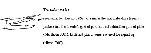
Peritremes are the integument at the edge of the undershell that surround the spiracles to be discussed later. As you can see, the genital plate is “arrowhead-shaped”, and appears only in females. The male would have a gonopore at the top of the sternal shield, and “spermadactyl fused to the movable cheliceral digits” (Knee and Proctor 2006). NFM has a tear-shaped anal plate (Weisbroth 1960).
I haven’t shown all the parts of the legs. Starting at the body and moving towards the claw, they are as follows: coxa, trochanter, femur, genu, tibia, and three tarsi, ending in a tarsal claw (Knee and Proctor 2006). Joints are necessary for movement with the hard outside coating. Interestingly, the multi-layered exoskeleton (or integument) that is so critical to mite survival, can also be an evolutionary size control, since the larger it is, the more energy it takes to support (Thanukos 2007). Hence the biggest-shelled arthropods live in the water (Audesirk et al. 2008).
The Internal Organs

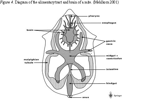
Mites may be tiny, but they have many of the same systems that we have: digestinal, excretory, nervous, and reproductive to name a few. Figure 4 shows some of the major organs. Liquid food comes in through the buccal cavity and pharynx, and down the esophagus to the midgut. Digestion is both intra- and extracellular (Mehlhorn 2001).
The gastric ceca (blind gut) store food for times of famine. The partially digested food passes through to the intestine and hindgut. The malpighian tubules are excretory in nature, and carry other bodily waste to the hindgut where it joins digestive waste, and is discharged through the anus (Nixon 2007).
How do they breathe? Generally through a pair of tracheal spiracles (respiratory orifices) behind the third or fourth coxae of legs (Mehlhorn 2001). Specifically, O. sylviarum has a stigma labeled behind leg #3 on one of Knee and Proctor’s (2006) SEMs. These open and close by use of special muscles depending on the level of CO2 (Cloudsley-Thompson 1968).
Oxygen enters the tracheae, “narrow, branching, chitinous tubes”, by diffusion (Savory 1964). Since mite “blood” does not carry oxygen, arthropod size is limited by the rate of oxygen diffusion (Thanukos 2007). The circulatory system is open, and the organs are directly bathed in a fluid called hemolymph (Nixon 2007).
I was not surprised to find out that the mite had a brain since it seems pretty smart about seeking out hosts. (Though its location below the esophagus is a bit strange.) In fact it has a reasonably well-developed central nervous system that has “allowed for the evolution of complex behavior” (Audesirk et al 2008). There are different sensory structures on the body surface that are important since they have no eyes (Nixon 2007). The most common are hair-like setae. This mite has a group of them on the upper part of the tarsus of the first leg that function as an olfactory organ (Savory 1964).
Scientists are still investigating mite anatomy using scanning electron microscopy. A team in Spain has examined a close relative of the NFM, the red chicken mite (Dermanyssus gallinae), and has hypothesized that the porous setae are used for smell and the grooved ones are used to detect mechanical stimuli (Soler Cruz et al. 2005). A study done in 2004 showed that the mites were very influenced by heat and vibration and use it for orientation (Owen and Mullens 2004). Other sensible areas used to detect chemicals (e.g. CO2) include the terminal segment of the palps and the cheliceral digits, the latter of which may sense heat (Sonenshine et al. 1986). Our exterminator told has that when he tapped and breathed on our light fixture, they came out in droves (Williams 2007 pers. com.).
In terms of reproduction systems, the mites are fairly sophisticated. The males have a gonopore at the end of an ejaculatory apparatus, a vas deferens, and testes. The females have ovaries, vaginas, and vaginal glands (Mathews Pound and Oliver 1976).

Ontogeny
Here are the stages of development, which can be completed in less than a week under perfect conditions: egg, larva, protonymph, deutonymph, and adult. After mating, the adult mites lay up to five eggs that hatch in about a day (Sikes and Chamberlain 1954). The larva only has three pairs of legs, but will have four as nymphs (Mehlhorn 2001). In less than a day, they molt into a protonymphs. After two blood feedings, they molt into non-feeding deutonymphs that then molt into an adults. Shedding the non-expanding shell is essential for growth, but is an evolutionary size limit for two reasons: invertebrates have no other support for the body, and it leaves the organism vulnerable to predators (Thanukos 2007). After another engorgement as adults, the adult mites lay their eggs and the cycle repeats (Sikes and Chamberlain 1954).
Adults can go for three to four months without a host (Clark 1991). If the population is smaller, the mites will spend their whole life on the birds (Sikes and Chamberlain 1954). If not, they alternate, spending more time in the nest when not feeding (Proctor and Owens 2000).
